
Treating craniosynostosis in infants has traditionally required complex surgical correction. But advances in minimally invasive craniofacial approaches have greatly improved outcomes and changed what families can expect from such procedures. Dr. Timothy Vogel has consistently advocated for the use of refined tools and gentler operative methods in treating the condition where the skull bones fuse too early. An internationally respected board-certified pediatric neurosurgeon, Dr. Vogel believes that these are safer and more efficient treatment options for babies diagnosed with craniosynostosis.
A newborn’s skull is meant to grow along flexible sutures. When one or more sutures fuse too early, the resulting pressure buildup can affect brain growth and development. Left untreated, craniosynostosis can limit neurological development and cause visible head shape differences, making early diagnosis essential. With recent advances in surgical planning, Dr. Vogel can treat patients with greater precision and minimize the impact on infants.
Each case is unique in its presentation, its impact on the family, and its complexity. Dr. Vogel shines in his compassion towards the infants and the families, and in his extensive experience and knowledge of complex conditions that affect children with craniosynostosis. His patients and families rave about him and his team’s comprehensive and individualized care. These factors help make Dr. Vogel and his staff a highly sought-after craniofacial team in the New York, New Jersey, Connecticut, and Pennsylvania areas. As a result of the time spent with each individual family, Dr. Vogel has become busy with his minimally invasive surgical care at the leading children’s hospitals.
The Value of Early Intervention
The first months of life are critical for babies with craniosynostosis, because their bones are so soft and bone growth occurs rapidly. However, minimally invasive endoscopic surgery can guide proper expansion with far less operative stress than traditional methods. This allows the brain to expand and take its normal shape without impacting development.
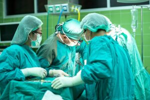
In endoscopic procedures, Dr. Vogel creates a small incision to release the fused suture and allow the skull to resume natural growth. Unlike open cranial vault reconstruction or springs, which traditionally required larger openings and more extensive remodeling, endoscopic surgery works with the infant’s own biology to gradually reshape the skull.
Dr. Vogel supports the continued advancement of these early interventions. He notes that technology has made these procedures safe and well tolerated by the infants, with just an overnight stay in the hospital, which results in timely treatment, shorter hospital stays, and reduced discomfort.
Endoscopic-assisted surgery improves visualization, which means surgical teams can now operate with greater clarity while minimizing blood loss. This has been one of the most significant improvements in infant craniofacial care.
Improved Imaging and Navigation
Occasionally, for complex cases, high-resolution imaging has become invaluable for diagnosing multiple suture craniosynostosis and for mapping the surgical route. In such complex cases, imaging allows for detailed three-dimensional scans, providing doctors with a clear view of fused sutures, skull curvature, and areas where pressure may affect brain development. These images help guide surgical strategy, allowing surgeons to identify the most efficient path to relieve the fusion.
During surgery, real-time endoscopic navigation further improves the accuracy of the procedure. The endoscope allows surgeons to maintain a precise orientation while working through extremely small openings. This control reduces the risk of harm to adjacent tissues and helps achieve consistent results across a range of craniosynostosis types.
In his experience as a pediatric craniofacial surgeon, Dr. Vogel cites the value of integrated planning tools in streamlining the operative process. He cites the benefits of digital modeling, particularly the use of preoperative scans to create virtual guides when needed for multiple suture craniosynostosis. This helps anticipate variations in skull shape and account for subtle differences across babies.
Endoscopic Surgery: What to Expect
Endoscopic surgery is particularly helpful for infants who are diagnosed early, offering a less disruptive alternative to traditional cranial vault remodeling. The procedure involves making a small incision, less than an inch in size, through which a small pen camera and instruments that release the fused suture are inserted. The minimal access required reduces swelling, shortens anesthesia time, and virtually eliminates the need for blood transfusion. Babies often recover quickly, returning home the next day. This is 3 to 4 days sooner than with open approaches.
Following surgery, patients are fitted with a helmet that aids in the reshaping process. Because the skull is still expanding rapidly, it is necessary to use a custom-fitted orthotic helmet that gently directs growth toward a balanced contour. Dr. Vogel helped to design the helmet that leading craniofacial centers now use across the United States and his team has a helmet orthotist who helps to care individually for each patient. Having this resource allows Dr. Vogel’s team to carefully monitor response to the helmet, make adjustments, and continue to check on his patients and families as they go through the helmet process, which may last 3-4 months.
These methods underscore the importance of early diagnosis. Infants older than 7 months of age usually require more extensive reconstruction because their skulls harden as they grow. By focusing on newborns through roughly the first 6 months of age, endoscopic treatment uses natural growth to its advantage and eliminates the impact on developmental delays.
Postoperative Care and Developmental Monitoring
Dr. Vogel notes that the postoperative period does not require any extensive special care. Infants are able to feed and be held right after surgery, and because the incisions are small, any discomfort is seen in just a few hours after surgery. Babies return to themselves hours after surgery and can go home the next morning. Following the hospital course, Dr. Vogel and his team see patients each week for the first 3 weeks to check on the infants, the incision, and the helmet. Dr. Vogel and his team are committed to seeing that these children have a bright future, and he continues to follow his patients yearly until the age of 7-8 years old to ensure that they thrive in their development.
A Clearer Path for Families
Craniosynostosis can be an overwhelming diagnosis for parents. However, the evolution of minimally invasive surgery is a significant leap forward in better developmental outcomes and early recovery.
Dr. Timothy Vogel continues to support advancements that lead to shorter recovery times and improved success rates. He continues to work on refining craniosynostosis surgery to ensure its safe and effective nature for infants. Dr. Vogel leads a highly skilled, busy, and compassionate team, and through his efforts, infants can grow with healthy development. Each family deserves individual care for their child. Dr. Vogel’s minimally invasive approaches are gentler and more effective procedures for newborns diagnosed with this condition.