
Much of the conversation about longevity focuses on what we might take, change, or invest in to live longer. I have become increasingly interested in an earlier question: how well are we identifying health problems while there is still an opportunity to act?
That question became personal when I tried a 3D breast ultrasound through BeSound. I had never had a mammogram. I had heard unsettling stories about the discomfort, and I had questions about what mammograms could miss.
Recently featured in Fortune and Forbes, BeSound was founded by a former NASA intern physicist, Bailey Renger, and designed alongside Lavinia Errico, co-founder of Equinox.
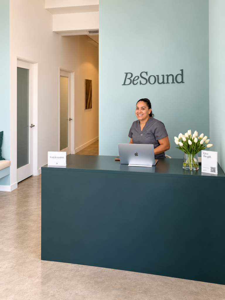
The women-centered design was immediately apparent when I stepped inside the West Hollywood location, with robin’s egg blue walls, soothing music, and fluffy spa robes.
My scan felt remarkably easy. It took about five minutes, and I received my results within half an hour.
A pleasant appointment cannot establish how well a technology detects cancer. But it can prompt a useful question: what would better screening look like if we considered both the clinical evidence and the person experiencing it?
Where AI Is Making a Measurable Difference
Breast imaging offers a concrete example of how artificial intelligence can support diagnosis. AI systems can flag suspicious areas on images and help determine which examinations need additional review. The important test is whether that support improves care in practice.
In the Swedish Mammography Screening with Artificial Intelligence trial, known as MASAI, researchers compared AI-supported screening with the standard approach of having two radiologists read mammograms. A 2025 analysis reported cancer detection rates of 6.4 per 1,000 participants with AI support, compared with 5.0 per 1,000 in the control group. That represented a 29% relative increase in detection, or approximately 1.4 additional cancers identified per 1,000 women screened. Screen-reading workload fell by 44.2%.
Those findings are encouraging, but their meaning matters. More cancers detected does not automatically establish fewer deaths or less intensive treatment. Those outcomes require follow-up, alongside attention to false alarms and overdiagnosis, when screening finds a cancer that would never have caused harm.
The opportunity is to help clinicians make better decisions with the time and information available. That is a more useful benchmark than whether a product simply includes AI.
Why Breast Screening Matters
The scale of breast cancer makes progress consequential. In 2026, an estimated 321,910 women in the United States will be diagnosed with invasive breast cancer, and 42,140 will die from the disease.
Outcomes vary substantially by how far the cancer has spread at diagnosis. National Cancer Institute data report five-year relative survival of approximately 100% for breast cancer confined to the breast, compared with 33.8% for cancer that has spread to distant parts of the body. These are population estimates, not individual guarantees, and the comparison alone does not prove the benefit of screening.
There is separate evidence from randomized trials that screening mammography reduces breast cancer deaths. The U.S. Preventive Services Task Force recommends mammograms every two years for women at average risk between ages 40 and 74. Screening plans may differ according to individual risk and the guidance a clinician follows.
Mammograms also have limitations. The American Cancer Society estimates that screening mammograms miss about one in eight breast cancers, with misses more likely in dense breasts. Abnormal results can lead to additional imaging or biopsies even when no cancer is present.
These tradeoffs deserve clear explanations. Early detection does not generally prevent breast cancer from developing. Its purpose is to find disease before symptoms appear, when treatment may have a better chance of helping.
A Broader Opportunity in Women’s Health
Breast screening also belongs within a larger conversation about women’s health. The NIH Office of Research on Women’s Health describes how the historical exclusion of women from clinical trials left gaps in the evidence used to guide their care. Understanding differences in symptoms, disease patterns, and treatment responses remains essential to delivering care tailored to women’s needs.
For me, that makes women’s health an important lens through which to evaluate diagnostic innovation. Are women appropriately represented in the studies? Does a technology perform well across different ages and patient groups? Are patients given clear explanations of its limitations and what happens after an abnormal result?
These questions matter for AI as much as for imaging devices. I would want evidence that a tool works for the women who will actually use it, alongside attention to comfort, cost, and access. A welcoming appointment is valuable. So is knowing that the decisions it informs rest on research that reflects your needs.
What I Experienced at BeSound
Against that background, I was curious about a different imaging experience. Stories about mammograms had shaped my expectations before I had ever undergone one myself. I wanted to understand what a newer approach felt like.
BeSound uses 3D automated breast ultrasound, specifically the GE ABUS, that is FDA approved for breast cancer screening. Ultrasound uses sound waves to produce images of breast tissue, rather than the X-rays used in mammography. The automated system captures images across the breast and assembles them into a three-dimensional view. A physician interprets the images.
A 3D breast ultrasound is also different from a “3D mammogram,” or digital breast tomosynthesis, which is an X-ray examination. The shared term “3D” can obscure that distinction.
They also have a second machine that is FDA approved
BeSound also has an Imagio Breast Imaging System. This is a second FDA-approved technology that combines ultrasound with optoacoustic imaging to provide more information about the metabolic activity of a particular region. Using pulses of laser light and ultrasound, it provides information about blood supply and oxygen levels in and near to a breast mass. This can help physicians distinguish benign and malignant findings without ionizing radiation. The optoacoustic imaging is only offered to screen lumps or masses identified during a breast work-up. It does not replace mammography or a biopsy when one is needed.
For me, the experience was pleasant and straightforward. The scan took about five minutes, and I had results within half an hour. Those timings describe my appointment: BeSound currently advertises approximately 20-minute visits and reports within 24 to 48 weekday hours.
The experience made me think about the emotional side of preventive care. When an appointment feels manageable, it can change how we feel about returning. That is my personal reflection, rather than evidence that a particular service improves screening participation.
For Bailey Renger, founder and CEO of BeSound, that return visit is also an opportunity to build a more complete picture of a woman’s breast health over time.
“Every BeSound scan captures a complete, standardized 3D image of the breast, and that consistency is what makes AI genuinely useful in breast imaging,” Renger told me. “We’re building the foundational AI layer that helps physicians read faster, spot subtle changes earlier, and follow a woman’s breast health over time instead of judging a single snapshot. AI doesn’t replace the physician. It can give them a sharper view, and it can give women answers sooner.”
The clinical role of ultrasound requires equal attention. In the SomoInsight study of 15,318 women with dense breasts, mammography detected 82 cancers. Adding automated breast ultrasound identified 30 additional cancers, approximately 37% more than mammography alone. In this study, that increased detection from 5.4 to 7.3 cancers per 1,000 women screened. The additional detection came with a tradeoff: more recalls for further evaluation.
Crucially, the study evaluated ultrasound alongside mammography. It did not establish ultrasound alone as a superior replacement. BeSound describes its imaging as supplemental and states that it does not replace mammography. My ultrasound experience therefore does not remove the need to establish an appropriate mammography screening plan with my clinician.
What Meaningful Progress Should Deliver
For Ariel Ganz, PhD, the promise of better diagnostics is deeply personal. In a MedCity News interview, she described how an elective MRI detected her breast cancer after earlier imaging had appeared normal.
That experience helped shape the investment philosophy behind Arben Ventures, which she co-founded to back scientific breakthroughs at the intersection of technology, medicine, and AI, with a vision to prevent, detect, and cure disease.
“I was diagnosed with breast cancer at 33, before routine screening begins and I feel incredibly fortunate that my cancer was detected early with elective imaging,” Ganz told me.
Her priorities for better screening include identifying who could benefit earlier and making appropriate imaging more accessible.
“Young women get cancer and deserve access to imaging. We need to identify women who could benefit from earlier screening and make the appropriate imaging easier to access.”
Ganz also points to gaps in access to supplemental screening, an issue that informed Arben’s investment in BeSound.
“For women with dense breasts, studies show that ultrasound can find cancers mammography misses, and still insurance coverage for supplemental screening is not equally available to all women and different women are being offered different care. That is part of why we invested in BeSound at Arben Ventures: to help close these gaps and make breast imaging more accessible.”
Her perspective brings the conversation back to a practical measure of progress: whether innovation helps women receive appropriate, evidence-based care at the right time.
My BeSound appointment gave me a positive experience of breast imaging and a reason to learn more. It also reminded me to distinguish how a test feels from what it has been proven to do.
As an investor and a patient, I want to see progress on both fronts: reliable detection, clear explanations, appropriate follow-up, and appointments that feel easier to approach. The promise of longer, healthier lives depends in part on how well we deliver those things today.